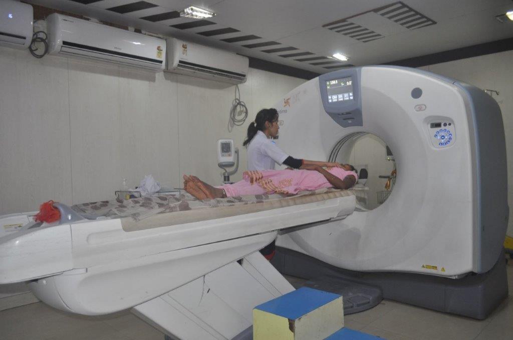
CT Scan
X-ray computed tomography (CT) is a medical imaging method employing tomography created by computer processing.

Since the advent of x-rays in the late 19th century and the technique of CT scanning was developed in the 1970's, a range of increasingly advanced imaging techniques mean that doctors today have a host of scanners at their disposal.
At Gupta Diagnostic Clinic, one the most technically advanced CT scanners on the market is available to our referrers and patients. Called the High Speed Dual Slice, manufactured by GE Healthcare, this remarkable machine is paving the way for more accurate diagnosis of diseases and early warnings of potential problems.
The HighSpeed VCT's groundbreaking technology opens the door to new procedures while improving the existing ones. Faster coverage with sub-millimeter resolution and shorter patient-breath-holds allow more robust and repeatable studies. This is critical in emerging procedures such as Coronary Angiographic studies.
What is a CT scan and how does a CT scanner work?
A CT (Computerised Tomography) scanner is a sophisticated machine that uses x-rays to acquire images of the body. Instead of sending out a single x-ray beam through your body as with ordinary x-ray examinations, a fan-shaped beam of x-rays passes through a slice of your body onto a bank of detectors where their strength is measured.
Beams that have passed through less dense tissue such as the lungs will be stronger, whereas beams that have passed through denser tissue such as bone will be weaker.
A powerful computer will use this information to work out the relative density of the tissues and the results are represented as a cross-sectional, two-dimensional picture shown on a monitor.
What are CT scans used for?
Any part of you body can be examined to assist in the diagnosis of your medical condition. CT scanning is particularly good at looking at the following areas:
- Internal organs within the chest and abdomen e.g. lungs, liver, spleen, pancreas and kidneys
- Orthopaedic imaging i.e. bones
- Brain imaging
- Vascular imaging to exam blood flow
CT scanning can also be used to guide biopsies and therapeutic pain procedures.
CT scans are far more detailed that ordinary x-rays. The information used to create the two-dimensional computer images can be reconstructed to produce three-dimensional images which can then be used to produce virtual images that show what a surgeon would see during an operation. CT scans can allow doctors to inspect the body without having to operate or perform unpleasant examinations.
Preparation for the scan
For some examinations, no preparation is required, However if the patient is having a scan of the abdomen, for example, they will be asked not to eat for up to 6 hours before the test. For many examinations of the abdomen, the patient will be given a drink containing gastrografin, an aniseed flavoured x-ray dye, up to an hour before the procedure. This makes the intestines easier to see on the images. In other examinations of the abdomen, the patient will be given a cup of water to drink immediately prior to the examination.
Metal can interfere with the image quality, so everyone who has a scan is asked to remove metal objects such as coins, jewellery, and hair clips, and it's best to wear clothing that does not have metal zips or buttons.
The scan
Having a CT scan involves lying on a table that slides through the 'ring' of the scanner. The table is positioned so the part of the body being examined lies within the ring. The table moves through the gantry as the x-ray source and the detectors rotate around inside. There will be a whirring noise caused by the moving parts of the scanner within the gantry.
The radiographer operates the scanner from behind a window, and is able to see, hear and speak to the person being scanned throughout the procedure. The patient will be asked to keep very still and hold their breath for a few seconds.
For some examinations, an injection of contrast agent (x-ray dye) is needed to enhance visualisation of organs and blood vessels. It is usually given into a vein in the arm or hand at the beginning of the scan.
The total length of a CT scan procedure can vary from between 10 minutes to an hour. The actual scanning itself takes between a few seconds and a few minutes depending upon the examination, so most of the procedure time is in preparation and confirmation that the images are of sufficient quality.
Once the examination is over most people can resume their normal activities immediately. However, if you have had an injection involving a local anaesthetic (usually in the neck or back) you will not be able to drive or operate machinery for the remainder of the day.
The images acquired will be interpreted by a radiologist and the results will be sent to the doctor who arranged the scan.
Deciding whether to have a CT scan
A CT scan is a commonly performed and safe procedure. There is very little that can go wrong. However, in order to give informed consent, all patients deciding whether or not to have this procedure need to be aware of the possible side effects and the risk of complications.
Side-effects
The CT scan itself does not have any physical side effects. However, if a contrast agent is used, some people may be affected.
As the dye is injected it can cause flushing and sometimes nausea. Some people feel warm and get a strange metallic taste in their mouth. These are normal sensations and pass within a few seconds.
Complications
The only complications that may arise from a CT scan are related to the contrast agent. Some people may have an allergic reaction to the dye. This is very rare and can be treated immediately with appropriate medicines. Also, the dye can cause further kidney damage in people who already have kidney problems.
People who have allergies or kidney problems should tell the CT department staff prior to the examination.
Contraindications
CT technology requires the use of ionising radiation. There known but slight risks to the foetus if exposed to ionising radiation, therefore if there is any chance that you could be pregnant, you will not be scanned.


